Lower Back Muscle Anatomy Diagram : constructive critisism. The lower trapezius, middle trapezius and upper. Muscle anatomy diagram labeled 12 photos of the muscle anatomy diagram labeled muscle anatomy diagram back, muscle anatomy diagram female, muscle anatomy diagram pdf, muscle anatomy diagram printable, muscle anatomy diagram quiz, human muscles. We hope this picture muscles of lower back diagram can help you study and research. It's a cylindrical muscle that travels along the length of the spine. Learn anatomical details of the lower back muscles, so you can draw them.
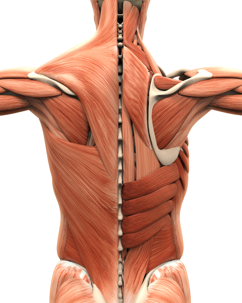
You can click the image to magnify if you cannot see clearly. First a few words about anatomy: Human muscles enable movement it is important to understand what they do in order to diagnose sports injuries and prescribe rehabilitation exercises. 16.06.2020 · anatomy of the lower back muscles diagram, diagram of lower back muscles pain, lower back muscles diagram pain The back anatomy includes the latissimus dorsi, trapezius, erector spinae, rhomboid, & teres major.
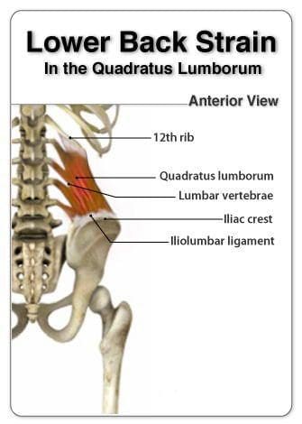
Quadratus Lumborum Pain | El Paso, TX Doctor Of Chiropractic from www.dralexjimenez.com For more anatomy content please follow us and visit our we think this is the most useful anatomy picture that you need. Muscles get their energy from different sources depending on the situation that the muscle is working in. The lumbar spine is the lower part of the back. Learn the lower back muscle anatomy associated with low back pain and hip pain. They are located deep to the extrinsic muscles, being separated from them by functional anatomy: Click on the labels below to find out more about your muscles. Human muscle system, the muscles of the human body that work the skeletal system, that are under voluntary control, and that the following sections provide a basic framework for the understanding of gross human muscular anatomy, with descriptions of the large muscle groups and their actions. The back comprises the spine and spinal nerves, as well as several different muscle groups.
The muscles of the spine anatomy chart shows every one of the many layers of muscle in the spine and back, using beautifully illustrated and detailed representations of the human.
The lumbar and sacrum region make up the bone of the lower back anatomy. Okay so this tutorial is on the back muscles. They are a gland, so there is a. Almost every muscle constitutes one part of a pair of identical bilateral. If you want to learn a bit more in detail about the back muscles, look at my individual tutorials on the extrinsic for some reason on this model, the splenius cervicis isn't shown, but this originates a little bit lower down and inserts onto the transverse process. The spinal cord is contained within the spine's vertebrae, running through the vertebral foramen and branching out to the peripheries through. The latissimus dorsi originates from the lower part. This image added by admin. There are around 640 skeletal muscles within the typical human body. Learn about anatomy back muscles with free interactive flashcards. Anatomical diagram showing a front view of muscles in the human body. More specifically, from the crest of the sacrum and the posterior. Occipital bone, ligamentum nuchae, spines of all cervical vert… upper fibers into lateral third of scapula;
Alle muscles are detailed described incl. Lower back muscles anatomy anatomy back anatomy bones gross anatomy human body anatomy muscle anatomy bones and there are anterior muscles diagrams and posterior muscles diagrams. Human muscle system, the muscles of the human body that work the skeletal system, that are under voluntary control, and that the following sections provide a basic framework for the understanding of gross human muscular anatomy, with descriptions of the large muscle groups and their actions. The back comprises the spine and spinal nerves, as well as several different muscle groups. Creatine phosphate donates its phosphate group to adp to turn it back into atp in order to provide extra energy to the muscle.
Muscle and ligament pain in the lower back from buxtonosteopathy.co.uk The spinal cord is contained within the spine's vertebrae, running through the vertebral foramen and branching out to the peripheries through. Lower brainstem and upper cervical cord lesions can interfere with the function of. It's a cylindrical muscle that travels along the length of the spine. Muscles that move the lower jaw. The back anatomy includes some of the most massive and functionally important muscles in the the traps consist of three sections of muscle fibers: Learn the lower back muscle anatomy associated with low back pain and hip pain. The lower trapezius, middle trapezius and upper. Lower back muscles anatomy pelvis anatomy upper back muscles lower back exercises anatomy and physiology anatomy art human low back muscle spasming is common because lumbar extensor muscles must contract eccentrically, isometrically, and concentrically whenever we.
Occipital bone, ligamentum nuchae, spines of all cervical vert… upper fibers into lateral third of scapula;
In anatomical terminology, chewing is called mastication. The latissimus dorsi originates from the lower part. Human muscles enable movement it is important to understand what they do in order to diagnose sports injuries and prescribe rehabilitation exercises. Human muscle system, the muscles of the human body that work the skeletal system, that are under voluntary control, and that the following sections provide a basic framework for the understanding of gross human muscular anatomy, with descriptions of the large muscle groups and their actions. If you want to learn a bit more in detail about the back muscles, look at my individual tutorials on the extrinsic for some reason on this model, the splenius cervicis isn't shown, but this originates a little bit lower down and inserts onto the transverse process. Patients with tight hamstrings tend to develop. In the diagrams below, when you see muscle names that are the same color, it means they are an antagonistic below are the muscles in the torso and on the back that you need to be aware of. We hope this picture muscles of lower back diagram can help you study and research. Muscle anatomy diagram labeled 12 photos of the muscle anatomy diagram labeled muscle anatomy diagram back, muscle anatomy diagram female, muscle anatomy diagram pdf, muscle anatomy diagram printable, muscle anatomy diagram quiz, human muscles. Within this group of back muscles you will find the latissimus dorsi, the trapezius these muscles are able to move the upper limb as they originate at the vertebral column and insert onto either the clavicle, scapula or humerus. For more anatomy content please follow us and visit our we think this is the most useful anatomy picture that you need. You'll gain an understanding of how these muscles move, where they attach, and other anatomical details that will help you draw the lower back. Occipital bone, ligamentum nuchae, spines of all cervical vert… upper fibers into lateral third of scapula;
You can click the image to magnify if you cannot see clearly. The intrinsic back muscles, which are also called true back muscles. Human muscle system, the muscles of the human body that work the skeletal system, that are under voluntary control, and that the following sections provide a basic framework for the understanding of gross human muscular anatomy, with descriptions of the large muscle groups and their actions. Another key structure in low back pain is the hamstring muscles, the large muscles in the back of the thighs. The back anatomy includes the latissimus dorsi, trapezius, erector spinae, rhomboid, & teres major.
Muscle Diagram Of The Female Body With Accurate Description Of The Most Important Muscles Front ... from media.istockphoto.com The back comprises the spine and spinal nerves, as well as several different muscle groups. It originates from the pelvis; These support most of the body's weight. Patients with tight hamstrings tend to develop. Learn anatomical details of the lower back muscles, so you can draw them. More specifically, from the crest of the sacrum and the posterior. Here the extrinsic back muscles are classified into logical subgroups to facilitate knowledge. The splenius muscles originate at the midline and run laterally and superiorly to their insertions.
Creatine phosphate donates its phosphate group to adp to turn it back into atp in order to provide extra energy to the muscle.
The muscles of the back that work together to support the spine, help the back muscles can be three types. Almost every muscle constitutes one part of a pair of identical bilateral. Muscles get their energy from different sources depending on the situation that the muscle is working in. More specifically, from the crest of the sacrum and the posterior. Back muscles are arranged in several layers, so they are divided into deep and superficial, which, in turn, are arranged in two layers. This article covers the anatomy of the superficial muscles of the back, including trapezius the superficial back muscles are covered by skin, subcutaneous connective tissue and a layer of fat. Occipital bone, ligamentum nuchae, spines of all cervical vert… upper fibers into lateral third of scapula; Here we explain the major muscles of the human body. They are located deep to the extrinsic muscles, being separated from them by functional anatomy: The splenius muscles originate at the midline and run laterally and superiorly to their insertions. If you want to learn a bit more in detail about the back muscles, look at my individual tutorials on the extrinsic for some reason on this model, the splenius cervicis isn't shown, but this originates a little bit lower down and inserts onto the transverse process. In anatomical terminology, chewing is called mastication. The intrinsic back muscles, which are also called true back muscles.
Occipital bone, ligamentum nuchae, spines of all cervical vert… upper fibers into lateral third of scapula; lower back muscle diag. Another key structure in low back pain is the hamstring muscles, the large muscles in the back of the thighs.
Share :
Post a Comment
for "Lower Back Muscle Anatomy Diagram : constructive critisism"


Post a Comment for "Lower Back Muscle Anatomy Diagram : constructive critisism"